Types of x-ray sensor
X-ray sensors come in various types, each designed for specific medical imaging applications. Choosing the right type depends on the nature of the examination and the requirements of the healthcare facility. Below are the categories based on their operating mode and detection mechanism.
Traditional X-ray film
Traditional X-ray film uses a film-based system. It is the oldest method, utilizing X-ray-sensitive film to create images. When exposed to X-rays, certain areas of the film darken while others remain clear, forming an image of the body structure. It is commonly used in many general imaging applications due to its simplicity and cost-effectiveness. However, it requires chemical processing, which can delay results.
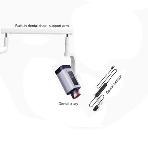
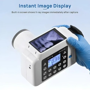
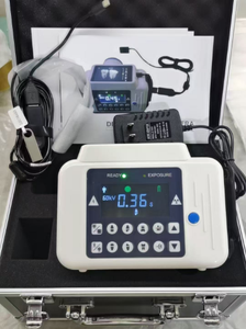
X-ray digital sensor
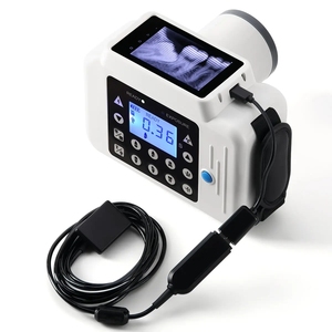
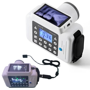
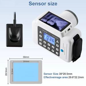
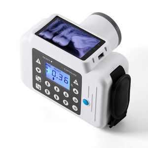
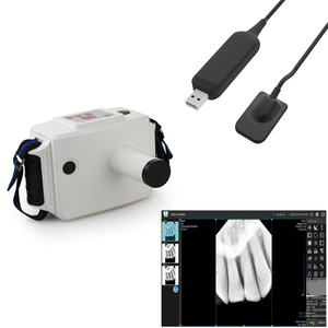
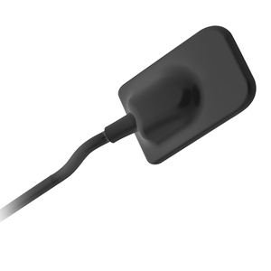
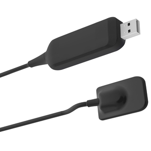
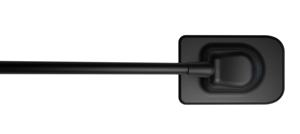
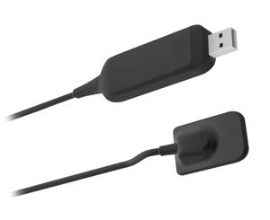
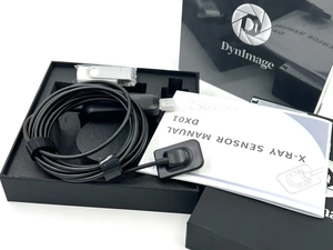
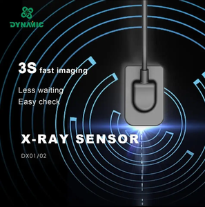
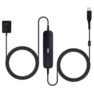
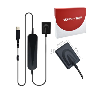
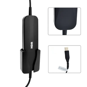
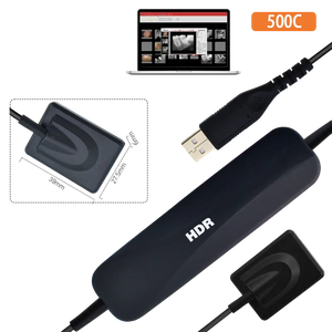
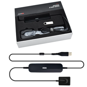
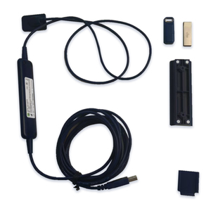
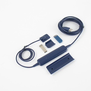
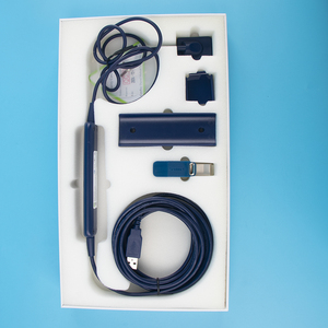
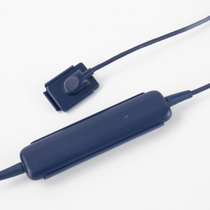
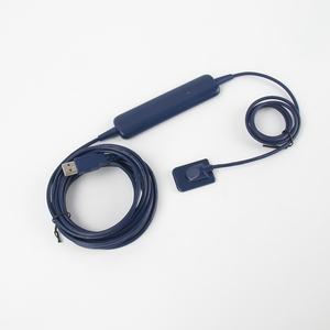
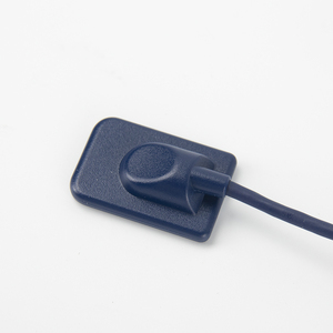
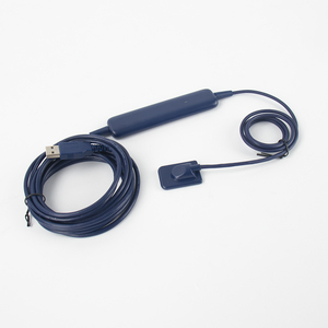
Digital X-ray sensors replace traditional film with digital sensors. It provides immediate images on a computer screen. It employs a flat panel detector (FPD) or a charge-coupled device (CCD) to capture the X-ray image. Immediate processing enhances efficiency in diagnosis and reduces radiation dose. It is largely used in dental practices, clinics, and hospitals.
X-ray imaging scanner
These are designed for specific applications, such as detecting foreign objects or ensuring quality control in industrial settings. It employs high-energy X-rays to penetrate dense materials. They are often used in security screening at airports and other high-risk venues.
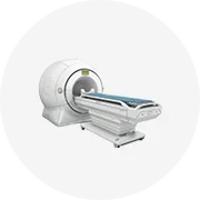
Computed Tomography (CT) scan
A CT scanner takes multiple X-ray images from different angles using a rotating X-ray source. It uses digital X-ray sensors (usually CCDs or amorphous selenium) to capture the images. CT provides cross-sectional images (slices) of the body, showing detailed internal structures in multiple planes. Industries widely use it in medical diagnostics, cancer detection, and trauma assessment.
X-ray CCD sensor
These sensors convert X-ray photons into electrical charges. It captures and digitally enhances X-ray images. The charges are then read out to form the image, allowing for high-resolution and fast image capture. In medicine, they are used in digital radiography and particularly in cardiac and cancer imaging.
Flat-panel detector (FPD)
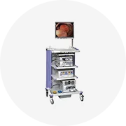
FPDs digitally capture an X-ray image by converting X-ray photons into visible light. In medical applications, they are used in digital radiography systems. These include in conventional and fluoroscopic imaging, where real-time video is essential, like in catheter placements or tumor biopsies.
Material & Durability of x-ray sensor
Maintenance of durability and image quality for the lifetime of an X-ray sensor requires such materials and construction considerations. Therefore, careful selection of the materials used to construct X-ray sensors is critical. This reflects their performance, durability, and application in various settings.
X-ray film sensor
Traditional X-ray films are made from a silver halide emulsion layer of sensitive crystals. These are typically gelatin-based films. It provides a balance between flexibility and resistance to tearing. The emulsion layer is coated onto a base layer of plastic or glass. This is to provide structural support. Once processed, the film's emulsion hardens, enhancing durability. However, films are sensitive to environmental factors like humidity and temperature. Proper storage in controlled conditions is vital for maintaining film integrity prior to use.
Digital X-ray sensor materials
Digital X-ray systems use different materials depending on the type of detector. CCD sensors for cat scans and other CT applications use silicon. They are known for their excellent image quality and light sensitivity. FPDs use scintillator layers. It consists of materials like cesium iodide (CsI) or gadolinium oxysulphide (GOS). These convert X-rays into visible light. The scintillator is coated onto a glass or polymer substrate that forms the base of the detector.
FPDs then use amorphous selenium (a-Se) or silicon (Si) to read the emitted light. Amorphous selenium is highly efficient at converting X-rays to electrical signals. Silicon sensors have high charges and can capture images with great resolution and accuracy.
Charge Coupled Device X-ray sensor
Conventional CCDs use silicon. It provides high image quality due to its efficient charge collection. The silicon matrix forms pixel arrays that capture electrical charges generated by X-ray exposure. X-rays then cause scintillation in a crystal. This produces light, which the silicon-sensitive CCD chip then captures. Silicon's hardness ensures stability. This makes it ideal for long-term use in medical imaging without degradation.
Inoganic scintillator
These are critical for converting X-rays into visible light. Common inorganic scintillator materials include sodium iodide (NaI), cesium iodide (CsI), and gadolinium oxysulphide (GOS). NaI is highly efficient but less durable than CsI or GOS. Therefore, they are often used in environments where the detector might experience rough handling or has high demands for radiation detection.
Commercial use cases of x-ray sensor
X-ray sensors are essential in healthcare and industry. This is for non-destructive testing, security screening, and imaging. There are various applications for which X-ray sensors find wide-ranging applications due to their ability to produce high-quality images of internal structures. Below are the scenarios in different contexts.
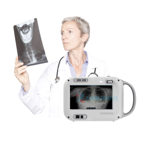
Medical applications
In medicine, X-ray sensors serve most imaging needs. They help in the diagnosis of such diseases as pneumonia, bone fractures, and cancer. It provides instant results. Their digital forms enhance efficiency by providing real-time imaging for procedures like catheter placements. Hospitals increasingly use digital X-ray systems to improve patient throughput and reduce the need for film processing.
X-ray scanning machines in the security industry
X-ray scanning machines utilize these sensors at airports and other secure venues. They detect contraband items and ensure public safety. Security personnel can identify threats such as weapons and explosives. The time for manual checks is reduced through this technology. Therefore, it enhances overall screening efficiency.
Industrial application
They increasingly use X-ray sensors for non-destructive testing (NDT). This is to examine components. In manufacturing, for instance, they inspect welds, solder joints, and other internal structures. This ensures product quality. X-ray machines also help detect density and voids in materials. Thus ensuring that only the best products reach the consumers. These sensors offer quick feedback to manufacturers. This allows for timely quality control, reducing the risk of defective products reaching the market.
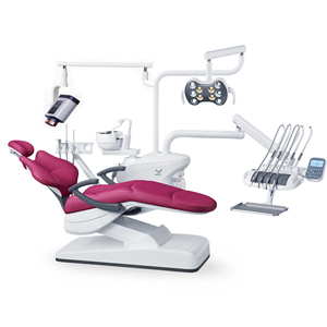
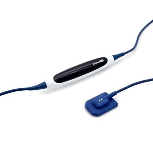
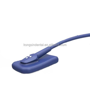
Dentistry
X-ray digital sensors enable easy dental diagnostics for practitioners. With instant radiographic images of teeth, bones, and surrounding tissues, they assist in cavity detection, periodontal disease, and other dental problems. Furthermore, the high sensitivity of these sensors reduces the amount of radiation required for patients. It makes dental clinics more efficient and improves patient care.
Research and veterinary applications
They leverage X-ray sensors to study the internal structure of animals. This helps them diagnose diseases and monitor health and injuries in pets and livestock. Research facilities also use X-ray imaging to study the effect of drugs and track the development of diseases in animal models and gain insights into new treatments.
How To Choose the x-ray sensor
Choosing the right X-ray sensor for customers depends on various factors. These are compatibility with existing systems, the specific applications required, and the budget to work with. They all play a key role in making the right choice. Below are more detailed factors to consider when selecting the X-ray machine for a use case.
Image resolution and sensitivity
Users need to ensure they get an X-ray sensor that has high image resolution. Also, they should get one that is very sensitive to the radiation it will receive. These two factors are very key. They determine how well the X-ray sensor will perform during medical imaging. Good resolution will allow the user to clearly view even the smallest structure that is in the human body. This will enable accurate he well and foster early diagnosis. Also, high sensitivity will minimize radiation exposure. This will also allow rapid image capture.
Type of application
Consider the environments in which the X-ray sensor will be used. In the medical industry, users will likely need digital radiography or fluoroscopy. In contrast, industries may require larger flat-panel detectors for CT scans. Also, high-volume screening will require high-throughput capability. This will ensure efficiency. Furthermore, for the dental industry, users should prioritize the digital sensor to take dental X-rays. This will capture high-quality images with very little exposure.
Digital X-ray sensor compatibility
X-ray sensors are not standalone devices that clients can use by themselves. Usually, they are integrated with other imaging devices. Users should ensure that the X-ray sensor they select is compatible with the existing devices of their customers. It will ensure seamless integration. For instance, hospitals may require digital radiography sensors compatible with computed radiography (CR) or direct radiography (DR) systems. Clinics, on the other hand, may need sensors that are simple to link to their current software for quick access.
Budget
It can not be easy to afford a good quality X-ray sensor. This is especially true for users who purchase them in bulk. Fortunately, they can get bulk discounts that will lower the overall cost. Nevertheless, it is still critical to consider the total cost of the sensor. That includes additional costs, such as maintenance fees, and running costs, like buying compatible equipment. Therefore, users should get X-ray sensors that offer them, the users, the best value for their money.
Q&A
Q1. What role do scintillator materials play in X-ray sensors?
Therefore, when choosing X-ray sensors for customers, it is vital to consider the performance of the scintillator materials. This is because they convert X-ray into visible light. This provides an essential step in the imaging process. High-quality scintillator materials will efficiently absorb X-rays and emit strong light. It will enhance image quality and sensitivity for X-ray equipment. This will help make accurate medical diagnoses and capture clear images. Thus, they will reduce radiation exposure to the patient.
Q2. Do digital X-ray sensors require special handling compared to traditional film?
No, digital X-ray sensors do not require special handling, unlike traditional films. This is because digital sensors do not need to be manually stored in a dark room or have chemical treatments applied to them, as is the case with film. The images will be almost instantly available for viewing with a digital sensor. It will speed up the diagnostic process and take up less space than traditional films. This makes them more efficient and easier to work with daily. That is why many healthcare facilities opt to get digital X-ray sensors over film sensors.
Q3. Are there any radiation risks associated with using X-ray sensors?
Fortunately, there are no radiation risks associated with using an X-ray sensor when it is used properly and with the required precautions. Also, digital X-ray sensors have been designed to minimize radiation exposure. They do this by efficiently capturing X-ray images. Furthermore, they allow immediate viewing. It facilitates quick feedback and reduces the total amount of radiation that a patient will be exposed to. Hospitals are advised to follow strict protocols for the maintenance and calibration of the sensors and imaging equipment. This will ensure optimal performance and minimal radiation exposure to the patients.
Q4. Which industries benefit most from using X-ray sensors?
Various medical and non-medical industries benefit from using them. These include hospitals for their various imaging needs and the dental clinic for dental radiography. Other industries have bulk and industrial X-ray sensors for non-destructive testing and quality control. Finally, high-security areas like airports use these sensors for screening and forensics.
Q5. How can one ensure the X-ray sensor chosen is durable enough for daily use?
To ensure the longevity of the X-ray sensor, its users should select one that is well-built and with proven materials. For example, those using scintillator coupled sensors should look for CCD sensors. It is because they have environmental protection and have resistance to wear and tear. Also, users should prioritize sensors that have robust housings. Finally, maintenance protocols should be established to ensure that the sensors remain in top condition even after heavy daily use.


![[ Aifan Dental ] Hot Sales <strong>Sensor</strong> Rayos <strong>X</strong> Dental I-<strong>sensor</strong> Digital RVG <strong>Sensor</strong> Dental](http://s.alicdn.com/@sc04/kf/H1ec70c513a5c4969b372b3c51c986ed7z.jpg_300x300.jpg)
![[ Aifan Dental ] Hot Sales <strong>Sensor</strong> Rayos <strong>X</strong> Dental I-<strong>sensor</strong> Digital RVG <strong>Sensor</strong> Dental](http://s.alicdn.com/@sc04/kf/H4123603a4bfe4f59aa70143eb17518eae.jpg_300x300.jpg)
![[ Aifan Dental ] Hot Sales <strong>Sensor</strong> Rayos <strong>X</strong> Dental I-<strong>sensor</strong> Digital RVG <strong>Sensor</strong> Dental](http://s.alicdn.com/@sc04/kf/H60583e729ccd4c0293c0e1aa6e1137bbP.jpg_300x300.jpg)
![[ Aifan Dental ] Hot Sales <strong>Sensor</strong> Rayos <strong>X</strong> Dental I-<strong>sensor</strong> Digital RVG <strong>Sensor</strong> Dental](http://s.alicdn.com/@sc04/kf/H800453d1614443babcf910899aeaab96J.jpg_300x300.jpg)
![[ Aifan Dental ] Hot Sales <strong>Sensor</strong> Rayos <strong>X</strong> Dental I-<strong>sensor</strong> Digital RVG <strong>Sensor</strong> Dental](http://s.alicdn.com/@sc04/kf/Haf8221d0634442999f01e0b0390dd2ffA.jpg_300x300.jpg)
![[ Aifan Dental ] Hot Sales <strong>Sensor</strong> Rayos <strong>X</strong> Dental I-<strong>sensor</strong> Digital RVG <strong>Sensor</strong> Dental](http://s.alicdn.com/@sc04/kf/He5c2c0968d904bcf86439b4851819a225.jpg_300x300.jpg)


 浙公网安备 33010002000092号
 浙B2-20120091-4