All categories
Featured selections
Trade Assurance
Buyer Central
Help Center
Get the app
Become a supplier
(1592 products available)


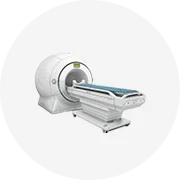
Animal imaging systems vary significantly. Higher-grade X-ray equipment can be useful for higher-density tissues and larger animals. Digital X-ray imaging systems are meant to enhance and expedite image acquisition and analysis.
Several of the most popular pet X-ray machine types include:
Digital X-ray machines
Digital radiology utilizes digital sensors instead of film, sending images to computers within seconds. Its picture adjustment features enhance diagnostics. Digital veterinary X-ray machines are more effective than traditional ones, providing vets with images quickly. The clearest shots reduce the need for retakes. Their sharpness highlights issues that may be missed on less sensitive equipment, giving a comprehensive view of pet health.
Portable X-ray machines
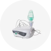
Portable X-ray machines are designed for animal care in locations with no facilities. They are lightweight and take batteries, so they're useful in emergency care or field situations. The wireless element means pets can be X-rayed outside a hospital. Field conditions do not affect image quality; portable machines provide digital precision in any setting. Quick imaging helps vets treat injured working or service animals away from base.
Fluoroscopy X-ray machines
Fluoroscopy is a real-time pet X-ray to view internal movement, such as in assessing swallowing or catheter placement. It gives immediate images rather than static, enabling live monitoring of internal processes. Fluoroscopy aids dynamic assessments that normal X-ray may not suffice. Watching in action helps diagnose issues with movement or function that can be missed standing still. It is a valuable tool to obtain information that otherwise would require surgery.
High-definition X-ray machines
High-definition X-ray machines boost clarity and detail, enabling better readings for difficult cases. These systems precisely image organs, bones, and foreign objects, giving complete assessments. Their accuracy helps identify small fractures or tumors. With such detail, these are standard in specialty clinics and emergency services. Their enhanced quality reduces retakes and increases the confidence level in accurate diagnoses.
The choice of a veterinary X-ray machine depends on several factors. One important consideration is the clinic's setup, which determines whether a digital or traditional system is more suitable. Exposure time is significant – quick imaging is vital for restless pets. For diverse patients, a portable machine must be compact and provide quality shots. Machines with advanced imaging software lessen the need for retakes by ensuring accurate initial captures. Other factors include budget considerations and available space.
Imaging requirements
Imaging requirements include the need for sensitivity to detect diseases and exposure time, which must be brief to avoid stress on pets. Larger animals need strong equipment, while small patients require detail. High definition is essential for spotting tiny issues like fractures or tumors. Special needs such as soft tissue imaging or live internal screening add to the machine's complexity. A suitable machine meets all these requirements for diverse patients.
Budget
The budget directly affects the veterinary facility's X-ray options. Radiography and digital setups cost different amounts, with digital typically more expensive. Financing plans and leases are helpful. Quality machines lower long-term costs by reducing retakes. Small clinics may afford a portable model instead of a fixed one. The best choice balances cost with effective diagnostics for quality care within budget limits.
Space available
The available space significantly impacts selecting the right pet X-ray machine. Digital systems are compact and may fit in a small area, while conventional machines require a more extensive space. Portable X-ray devices are even more space-efficient for smaller practices. Large veterinary hospitals may handle sophisticated machines that integrate with other imaging setups. The clinic's overall layout and whether renovations to install equipment should be considered.
Veterinary needs and future expansion
Considering future expansion is vital to meet the clinic's future needs. If one imagines a larger clientele or new services, an expandable machine with upgrade options is essential. High-capacity systems will suit busy facilities needing quick imaging without delays. Specialized features for advanced diagnostics will support future growth. A scalable investment avoids equipment changes with success and provides consistent service even as demands grow.
Veterinary X-ray machines help see problems inside pets without surgery. Doctors choose models based on the kinds of animals they treat the most. Regular maintenance makes the machine last. Keeping images secure helps patients and doctors work together. Here are some usage scenarios for portable x-ray machines for pets:
Routine checkups
Taking pictures during checkups helps find untreated problems early. Pet X-ray machines are valuable tools in routine healthcare. They help veterinarians internal checkups for hidden issues, taking pressure off the need for invasive exams. Routine imaging allows the discovery of growths, bone fractures, or joint dilemmas during standard visits. This proactive approach provides timely detection and treatment. Daily checkups enable vets to monitor ongoing conditions and make necessary adjustments to succeed in important health commitments.
Trauma cases
After accidents, X-rays quickly show breaks and damage. These scans allow timely, critical assessment of injuries. First help emerges faster with clear images for proper healing. Trauma situations require efficient diagnosis, and X-rays provide fast imaging results. By revealing fractures and internal issues immediately, they assist doctors in acting promptly and accurately.
Foreign body ingestion
Pets may eat things they shouldn't. X-rays help find where objects like sticks, socks, or toys are stuck. This internal view shows any blockages or danger, helping vets treat the problem sooner. Pets give themselves a tough time by eating the wrong stuff. When they do, vet techs rely on veterinary X-ray machines to locate the intruders. The pictures reveal any blockages or trouble caused by the items.
Pre-surgical assessment
X-rays taken before surgery help doctors plan better. They show if any surgery risks need more care, like issues with bones or inside parts. Viewing these images first leads to safer operations. Getting a clear plan means fewer surprises. The pet's health remains stable because the scans ensure that no vital change occurs before proceeding with surgery.
Assessment of chronic conditions
Pets with ongoing health problems like arthritis benefit from occasional X-rays. These scans check for changes over time, allowing better treatment. They keep track of how conditions develop. For pets with long-term illnesses, regular imaging shows changes that may impact treatment. It lets vets adjust the care plan according to each pet's current needs, ensuring optimum health management.
Proper maintenance is vital for keeping pet radiography machines working well. Simple tasks like checking the power source and cords or cleaning parts may be done regularly. Follow the machine's guide to schedule professional checkups. During busy times, repairs may be needed, so having a backup plan is wise. One might rely on a different unit or a portable X-ray. Quick replacement parts help keep work moving. Below are ways to ensure the pet X-ray machines stay healthy:
Routine maintenance
Pet X-ray machines work best when cared for properly. Regular checks and upkeep keep them running strong. Basic tasks, like cleaning filters and checking settings, should be done often. Bigger work, like calibrating parts, must be done by experts. Following the user's guide tells when to do basic care and repairs. Maintenance stops small problems before they grow big and keeps machines serving well for many years.
Common issues and troubleshooting
Poor images may come from dirty sensors or loose connections. Cleaning parts fixes many problems. Changing worn filters or tight belts may help, too. Sensors should be checked often for issues. Power problems are fixed by looking at wires and fuses. Referring to the user guide stops minor problems from slowing work. Users should learn to solve some of the small problems to avoid delays in working. Understanding alerts and errors reduces downtime. Digital machines sometimes need quick reboots if they freeze.
Professional repair services
Some repairs must be done by trained experts. They should service the machine when it breaks down badly or doesn't respond to basic care. Professional techs are needed for internal repairs, calibration, or software issues. Choosing an expert who knows the specific machine helps get it fixed faster. In multi-animal hospitals, repairs are started quickly to avoid delays in treating patients.
Part replacement
Parts like tubes, cables, and sensors will need changing over time. Backup parts should always be on hand. Computers link with X-ray machines, but if one breaks, the other can still work. Replacing parts quickly keeps work moving. Using only real parts from the manufacturer protects the warranty. Quick repairs and replacements keep the machine working well so that it may serve for many years.
A1. Digital imaging uses sensors instead of film, producing instant, high-quality images. This method enhances diagnostic capabilities with advanced software for quick, detailed views of X-rays. Its clarity assists better in finding issues. Unlike film, digital storage allows easy retrieval and sharing of images. This method is more efficient, with less time spent and more precise results.
A2. Usually, anesthesia for pets isn't needed during an X-ray. Most scans take only a moment, and many animals stay calm for the procedure. Though rarely, some restless pets may need light sedation to hold still. Anesthesia is avoided when possible to ensure quicker recovery. Vets always weigh the need for sedation based on the individual pet and the complexity of the scan.
A3. With portable machines, quick imaging types are done, like trauma X-rays or pre-surgical checks. They assist vets with emergency cases right at the site. These machines also work well for routine imaging in small clinics. Larger hospitals may also use them for imaging when another machine is busy. Their mobility and speed make them fit for diverse daily tasks, from simple to complex.
A4. Yes, special care is required because they deal with varied sizes and types. These precautions ensure accuracy and longevity. Regular cleaning, special calibration, and monitoring quality are needed due to this wide range of use. Tracking pet doses meets rules protecting from excess radiation. Specific tasks help maintain machines, keeping them ready for everyday demands.
A5. Secure storage protects shared and accessed images. Special systems store these safely, restricting who may view them. Encryption protects data from others trying to access it. Access controls ensure only certain workers see personal images. Digital rights management stops misuse. Auditing tracks all handling of these records. Together, these steps keep the images confidential.