All categories
Featured selections
Trade Assurance
Buyer Central
Help Center
Get the app
Become a supplier
(187 products available)


Suppliers often provide these types of MRI medical imaging equipment to buyers in bulk.
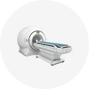
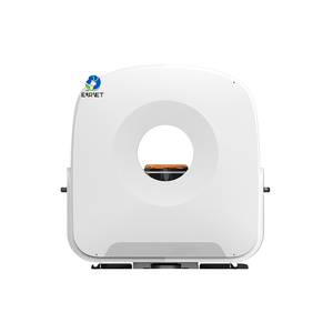
This is the most common type of MRI scanner used in hospitals and clinics. It has a large, cylindrical magnet that produces strong magnetic fields. This generates highly detailed images of organs and tissues. The closed MRI system provides the best-quality images. However, it has a narrow tube-shaped chamber which some patients find claustrophobic.
An open MRI scanner has a more open design. It allows patients to feel less confined during the procedure. It uses weaker magnetic fields than closed MRIs. However, it is not suitable for every patient. The images produced also tend to be of lower quality. Nevertheless, open MRIs are ideal for patients who experience anxiety in closed spaces.
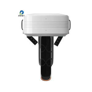
These closed MRI machines are designed to scan only the extremities. These include arms, hands, knees, and feet. Patients sit beside the machine while placing their limb inside the open-ended magnet. This type of MRI equipment is more compact and portable. It can be used in clinics or mobile units. It offers a more comfortable experience for patients who need scans of their extremities only.
This type of MRI machine is used primarily in research settings. It measures brain activity by detecting changes in blood flow. It helps researchers study brain functions, map neural pathways, and understand how different areas of the brain respond to stimuli.
MRA machines are used to create detailed images of blood vessels and arteries. It helps diagnose conditions like aneurysms, blockages, and vascular malformations. It uses specialized MRI techniques to visualize blood flow without the need for ionizing radiation.
These are high-resolution imaging machines used in hospitals. It uses a 3 Tesla magnet to produce faster scans with clearer images. The higher power magnet is ideal for detecting tumors, brain disorders, and joint problems. However, these machines are more expensive and require careful screening of patients.
Industrial application relies on these medical imaging machines.
They use MRI for drug development by studying how compounds interact with the body. It also helps visualize the effects of drugs on targeted organs or tissues. This provides detailed information about their efficacy and potential side effects.
These manufacturers of devices like contrast agents, coils, and catheters use MRI scanning. They use it to test and refine their products before going to market. It is also a quality assurance tool that helps ensure devices work effectively in real clinical settings.
They rely on MRI medical imaging to assess claims related to injuries, illnesses, and more. By examining the scans that correspond to a claim, insurers can better understand the nature of the medical condition being claimed.
In forensic cases, fMRI can be used to study brain activity related to memory and consciousness. This helps determine if someone was aware during a traumatic event. It, therefore, helps with liability cases and compensation determination.
These institutions use medical imaging in various fields of study. They are vital in neuroscience, where researchers use fMRI to study brain functions. In anatomy studies, MRI scans help create detailed maps of body parts for educational purposes.
Some factories have MRI machines to inspect innovative materials. That is how they evaluate the safety and effectiveness of new medical technologies. They test these machines before they are distributed to clinics and hospitals.
Here, the features and specifications of MRI equipment are highlighted.
Magnetic strength
The magnet strength impacts the quality of the images. Closed MRIs use a 1.5 to 3 Tesla (T) magnet. A 3T MRI enables the production of detailed, high-resolution images that are ideal for detecting small tumors, evaluating brain disorders, and imaging joints.
Open MRIs use lower power magnets around 0.3T to accommodate patients who experience claustrophobia.
Tesla
It refers to the strength of the magnetic field used in the MRI machine. Higher Tesla numbers indicate stronger magnets which can produce clearer and more detailed images. Most hospital MRI machines use a range of 1.5 to 3T.
Coil types
They are the components that help capture images during the scanning process. Different coils are used depending on what part of the body is being scanned. These include knee coils for imaging the knee and cardiac coils for the heart.
Show speed
The software analyzes the MRI scans to produce pictures of organs and tissues. Algorithms process the data quickly to generate images efficiently. Fast software speeds up the scanning process which is helpful in busy medical facilities.
Display
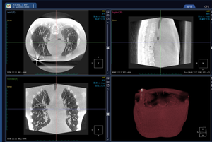
The display used should be high-resolution so that technicians can better view the scans. Most MRI machines come with large monitors to make the images easier to analyze.
Comfort features
Comfort is vital to alleviate patient anxiety. Open MRIs provide a less restricted area for scanning. Closed MRIs have wider openings and padded cushions that provide a more relaxing experience for patients during their scans.
Work with experts
It is vital for medical facilities to consult with their radiology colleagues about the best site for the MRI machine. They should hire experienced contractors in MRI installation as they know the best practices and are well aware of safety standards.
Assess power needs
Since MRIs are powerful machines, it is important to analyze whether the operating area has the right electric supply to keep the device functioning. They should verify that they have sufficient voltage and circuits to power the machine.
Prepare the area
Before the installation day, the medical facility should ensure that the MRI machine room is ready. The area should, however, be well protected to prevent dirt from gathering on important components during the installation work.
Schedule delivery
Before the installation process begins, the facility should arrange a delivery of the MRI scan so it can arrive on time. They should work together with the installers to plan for any barriers to getting the machine inside its designated space.
Prepare the patient
Before the imaging begins, the technician or technologist will ask the patient a series of questions to evaluate if they are fit for the MRI scan. They will provide the patient with ear protection to block out the loud noises the machine makes as it scans.
Position the patient
Patients are then instructed to lie on the scanning bed of the machine in a comfortable position. For patients with anxiety in confined places, techniques to minimize discomfort are applied.
Start the scan
The scanning process starts after positioning the patients. The bed slowly moves into the magnet tunnel of the machine. Strong magnetic fields then create images of the organs and tissues.
Provide postoperative care
Once the imaging is done, the technologist will help the patient get off the scanning table. The patient is then given time to relax if they feel any pain or discomfort. The technologist will afterward review the images to ensure the scan is of the required quality before sending it to the doctor.
Daily cleaning
The areas around the magnet and superconducting coil need to be wiped down with a nonabrasive, damp cloth on a daily basis. Radiologists should refrain from using cleaning chemicals that possess a high level of bleach.
Routine inspection
A technician should carry out an inspection of the scan machine's coil covers, cables, and transfer. They should inspect them closely for any indications of harm or wear.
Monitoring performance
Healthcare professionals should work with physicists to analyze whether the MRI's functions remain optimal. They should pay special attention to the quality of images produced and fluctuations in the device's performance, so a maintenance routine for those issues is established.
Check software updates
The MRI imaging software should always have recent updates installed. It maintains accuracy and boosts efficiency. The radiology department should work with the manufacturer to verify the software is updated.
Service by expert
After using the MRI equipment for maintenance and repairs, medical practitioners should always outsource professional help. They should avoid fixing the machine themselves even if they have seen tutorials online.
These are the key quality and safety requirements for these machines.
Strong magnets create better images, while weak open MRIs make lower-quality scans. Ensure the MRI machine has powerful magnets like a 3T that can handle complex scans. It guarantees detailed images without graininess.
Good software speeds up scans, and smart coils reduce distortion in images. Look for things like motion correction that avoid blurry pictures. Advanced software and hardware ensure the MRI has clear rather than foggy images.
The large magnetic fields used in MRI scans exert strong forces on metallic objects. They can even damage or displace non-removable implants surgically embedded in patients. To avoid these dangers, medical personnel should screen patients for metal fragments, shrapnel, or devices that may pose a risk when exposed to magnetic fields. They should also ensure all implanted medical devices, such as pacemakers or stents, are MRI-safe before scanning.]
Radiology departments should always have a safety checklist handy to verify patients present with none of the metals. They should include questions on dental work, piercings, and tattoos.
MRIs, unlike CT scans and X-rays, use magnetic fields to create images. They, therefore, produce no ionizing radiation. This makes them the preferred choice for imaging during pregnancy and for children. While performing an MRI, practitioners should be alert in case the patient reveals they are pregnant so they can take the right steps.
While MRI scans don't expose patients to radiation, some procedures use contrast agents. Hospital staff should watch patients closely for allergic responses after giving them these substances. They should know the signs of anaphylaxis and be ready to treat it, in case allergy happens.
Most patients experience a lot of stress during imaging because of the size of the machine. It is the technologist's duty to comfort them before and during the scan. They should also be alert to indications of pain or distress. They should offer reassurance and time for breaks if any discomfort occurs.
No, MRIs do not use ionizing radiation. They rely on powerful magnets and radio waves to produce images. This makes them safer than methods like X-rays, which do use radiation. However, always check with the doctor about the best method for individual needs.
An MRI provides a detailed look at soft tissues. It captures high-quality images of the brain, heart, liver, and joints. The scanning system can also examine organs like the pituitary gland, spleen, and uterus. Unlike other methods, MRIs excel at visualizing complex structures, making them useful for many medical assessments.
As long as they have no metal inside and feel okay during the scan, they can have as many MRIs as their doctor recommends. MRIs are one of the safest ways to see inside the body, and there is no limit to the number of times someone can get an MRI.
An fMRI measures brain activity. It creates detailed images using changes in blood flow to show which parts of the brain are working at any given time. It helps researchers map brain functions and understand how the brain responds to different tasks or stimuli. It is non-invasive and provides real-time insights into brain activity.
An MRI scan is useful for examining soft tissues, joints, and organs. It helps diagnose tumors, brain disorders, joint issues, and heart problems. It clearly shows the spine, liver, and pituitary gland. Its ability to capture high-quality images makes it valuable for many medical conditions, particularly those affecting complex structures.