All categories
Featured selections
Trade Assurance
Buyer Central
Help Center
Get the app
Become a supplier
(2161 products available)


 Ready to Ship


 Ready to Ship
 Ready to Ship


 Ready to Ship


 Ready to Ship


 Ready to Ship
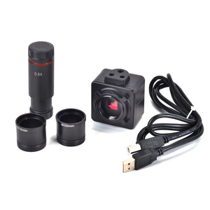
 Ready to Ship
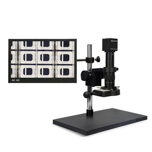
The eyepiece camera for microscope is categorised chiefly by its optical system, driving mode, and consolidation with various microscope types.
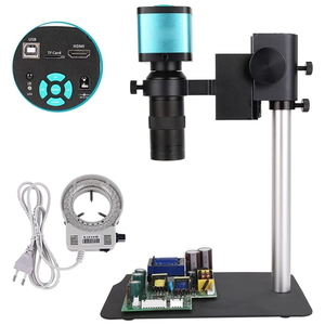
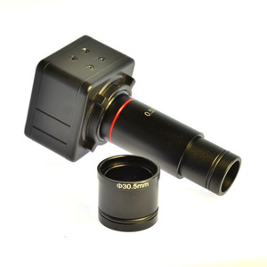
DIGITAL EYEPIECE CAMERAS
Such mainstream epics cameras for microscopes focus on capturing images using a computer or other digital device to view the images. An embedded image sensor and a data processing unit are used to collect and transform light rays, producing a digital image. Analysing these images online is truly convenient because of these cameras. Optical microscopes can send images via USB or Wi-Fi. No requirement for films; the images are emailed or posted quickly. Excellent for making, storing, and communicating images. These eyepiece cameras for digital microscopes fit most microscope types, including stereo ones.
ANALOG EYEPIECE CAMERAS
Before digital cameras took over, most people used analogue epipolar cameras. They take images on photographic film, though some systems employ analogue cameras to capture images on video, which is then transferred to the computer. Even though they're an outdated relic, some people still use them. Optical microscopy images captured by analogue epipolar cameras can be clear and sharp, especially when used on high-powered microscopes. These cameras connect to the microscope's eyepiece tube, though additional conversion lenses may be required depending on the type of camera and microscope model. Such cameras are more common in old labs and centres still using film for their microscopy needs.
EYEPIECE LASER CAMERAS
Microscopes for cell culture and research often use laser cameras to capture clear cell images. Compared to earlier cameras, these capture pictures far more intelligently by clarifying any blurry ones. A notable nature feature allows researchers to study microorganisms from a distance using a laser camera. Because of the enhanced resolution, further analysis is enabled for minute details on tiny structures. Its non-intrusive design makes it ideal for many lab environments requiring small subcellular components to be observed.
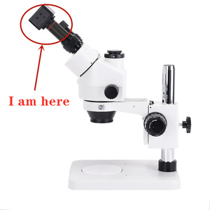
STEREOMICROSCOPE EYPIECE CAMERAS
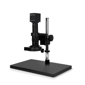
Stereomicroscopes are suited to handle 3D images of objects under study, as seen with the naked eye. These cameras are intended to capture patterns, texture, and structure information in research. They are mainly used in fields such as biological surveys and pinpointing defects in manufacturing. These cameras can deliver pictures with depth perception that allows further inspection in studies where physical structure is critical. The camera’s solid attachment to a zoom or stereo microscope makes it a convenient 3D imaging tool in microscopy.
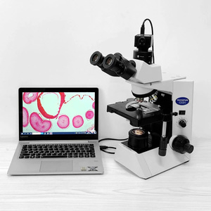
Most important: The role of an eyepiece microscope camera is to capture still and motion pictures of things through a microscope. Coupling the camera to the eyepiece of a microscope additions an extra lens to the system, which makes an image accessible on the camera sensor. Digital varieties can send images to computers for further manipulation. In education, pathology, and materials science, these cameras have become obligatory for documenting and examining microscopic subjects.
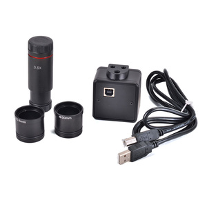
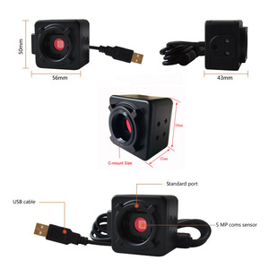
The design of eyepiece cameras concentrates on convenience and universality. Most digital devices consist of a camera sensor, an optical adapter, and a USB or Wi-Fi connection under a compact form factor. Software systems enable image capture and processing while simplifying the integration process with existing microscopes. Analogue cameras may be in a different style but are equipped with a more complex design that includes film or video capture. Stereomicroscopes use dual optics to produce 3D images, increasing depth perception in the camera images.
Situations for using an eyepiece camera for a microscope are variable, stretching across different fields where seeing microscopic items is needed. Such a tool can be found in various task environments, from academic research to medical diagnosis and industrial inspection.
BIOLOGY AND MEDICINE
Microscope cameras are important instruments in biology and medicine, where there is time and need to examine cells, tissues, and microorganisms. In histopathology, for instance, digital cameras allow pathologists to record and represent tissue slides for analysis and diagnosis. In microbiology, fish cameras are used to study bacteria and other minute organisms, bringing an image of their structure and development. Live imaging of cells is vital for understanding processes such as cancer development and immune responses. Digital eyepiece cameras help to document these processes for future reference.
MATERIALS SCIENCE
Microscopic cameras are used in materials science to study the microstructure of metals, polymers, and other materials. Electron microscopy cameras allow researchers to capture high-resolution images that reveal dislocations, grain boundaries, and phase distribution in a new material. These images are majorly important in improving material scientists. For more practical evaluations, digital cameras are used to capture defects in materials like fracture surfaces or inclusions under a stereo microscope. These evaluations are important for establishing material performance in engineering.
EDUCATION
Microscope cameras are popularly used in academic research. These instruments enable students to observe cellular structures, tissues, and microorganisms in biological science classes. The integration of digital cameras in teaching gives a chance for interactive learning, allowing students to capture and represent images for group study. This interactive method assists students in grasping microscopic concepts and discoveries better. In geology, they may study mineral structures and rock thin sections, and in industrial inspections, they may spot flaws or irregularities in materials.
INDUSTRIAL INSPECTION
In quality control and electronic manufacturing, eyepiece cameras for stereo microscopes enable technicians to pinpoint defects. The depth of field or 3D imaging capability in stereo microscopy makes it easier to check solder joints, circuit board assemblies, and intricate mechanical parts. Such visualisation allows for precision in multi-layered systems.
FORENSIC SCIENCE
Eyepiece cameras for microscopes are also valuable in forensic investigation. For instance, they assist in examining fibres, hair, and other trace evidence magnified under a microscope. Foreeic imaging combines pathology and engineering using digital cameras to capture and represent evidence in court.
The selection of an eyepiece camera for a microscope is determined by its purpose of use, the microscope type, and the attributes needed for effective imaging. Below are some guidelines.
Compatibility with MICROSCOPE
The camera must match its parent microscope to achieve the desired effect. For digital and other types of microscopes, a camera with a USB or Wi-Fi connection can be easily and quickly attached. Stereo microscopes need 3D imaging capabilities to perform better while examining objects under scrutiny. When dealing with high-power situations, an eyepiece camera must be coherent with an eclipse or other high-power microscope to maintain clarity when viewing small objects.
RESOLUTION AND SENSOR SIZE
Zoom in. Go big or go home, as they say. A camera with a higher resolution will be able to capture finer details of the sample. Cameras with resolution starting from 5 megapixels and above are ideal for materials science and forensic investigations. The size of the sensor is also important, as larger sensors capture more light and prove better in low-light conditions, which are so typical in microscopy.
Live Imaging AND FRAME RATE
Live imaging may be required, especially in fields such as biology and medicine, where changes occur in real-time. A camera with a good frame rate will give smooth live imaging. This is important in live cell imaging or when observing dynamic processes. Imaging rate is important when observing quickly moving live samples like protozoa or live insects. Choose a rate that matches the nature of the sample being studied.
SOFTWARE AND IMAGE CAPTURE
It is necessary to ensure the camera's software is compatible with the operating systems more likely to be used. The software system should put some functions and options in place for the processing of images, such as capturing, annotating, and manipulating them. This is especially important for labs where documentation-quality images are necessary. Industrial inspectors, educators, and forensic scientists also take drastic measures to ensure that quality images can be obtained controversial defects or evidence.
BUDGET
Eyepiece cameras for microscopes range from affordable to expensive, depending on specifications and uses. Digital models offering greater resolution and advanced software might be a little more appropriate for use in professional situations than their analogue counterparts. Budget makes a difference. Often, people switch from working with simple stereo or digital microscopes to using a high-end camera.
A1: Most digital eyepiece cameras fit excellently with light, stereo, and electron microscopes. It is always advisable to check if the camera is compatible with the specific microscope model, particularly when using analogue cameras.
A2: Stylistic factors greatly affect the price of an eyepiece camera for microscopes, such as the kind of camera, the power of the resolution, and extra features included. In other words, digital cameras of higher quality that may be used in more-lit professional environments could cost quite a lot, while simple models suitable for education or home use may cost significantly less.
A3: Yes. Digital epise cameras usually have imaging capture software bundled with them. This software should ideally run on commonly used operating systems such as Windows and macOS. These programs or packages enable users to capture and process images for documentation or analysis.
A4: Yes, many of these cameras come with high frame rates, which means they can give live imaging, especially to objects that are moving or changing in real time. Live cell imaging is greatly aided by this.
A5: One must consider the purpose of use, the type of microscope, and the desired specifications. Select a camera that is resolution, software, frame rate, and sensor size compatible with the requirements of the specific microscopy project.