All categories
Featured selections
Trade Assurance
Buyer Central
Help Center
Get the app
Become a supplier
(837 products available)


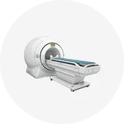
The echo color Doppler is a type of medical ultrasound equipment that shows blood movement through color imaging. There are a few major types based on imaging and blood flow study approaches.
2D Echo Color Doppler
The 2D echo color Doppler adds color to the standard 2D images to show how blood moves through the heart and vessels. While 2D gives a clear picture of the heart's shape and function, adding color shows the direction and speed of blood flow. This helps doctors understand conditions like valve problems or blockages. Most ultrasound machines used for routine heart checks at hospitals will perform this basic 2D echo color Doppler examination.
Pulsed Wave Doppler
This type measures blood flow speed at specific spots. The Pulsed Wave Doppler gets detailed flow speed data from one small area, allowing precise measurement of speeds right where the probe is focused. This contrasts with Continuous Wave, which surveys larger areas but gives less spatial detail. The detailed speed profiles help doctors spot narrowings or blockages by tracking slowed or abnormal flow in vessels feeding the heart.
Live 3D Echo Color Doppler
The innovative live three-dimensional technique enables doctors to view real-time volumetric color flow images around the heart. Unlike traditional two-dimensional echocardiography that provides flat cross-sectional pictures, the 3D method captures the anatomy in full stereoscopic vision. This breakthrough permits more accurate assessments of complex structures like heart chambers and vessel patterns. The fully spatialized shape conveys much richer information than 2D images, aiding precise surgical planning and intervention.
Continuous Wave Doppler
Continuous Wave (CW) Doppler is a highly sensitive method that measures blood flow speeds without limit, making it ideal for tracking fast flows in critical areas. By continuously emitting and receiving ultrasound waves, it captures the speed of blood even where flow rates approach peak velocities. This powerful capability outstrips other Doppler forms, allowing doctors to detect narrowing or obstruction signs in major vessels right to the heart. The detailed speed maps are crucial for diagnosing blockages and guiding treatment.
Assessment of Blood Flow
The primary task of the echo color Doppler is to visualize how blood moves through the heart and blood vessels. By superimposing color onto gray-scale images, it illuminates patterns of flow with clarity. Doctors can then see whether blood is flowing normally or if there are signs of obstruction, reflux, or other issues. This foundational function is key for routine checks and for diagnosing diseases.
Measurement of Flow Velocity
Beyond simple visualization, the Doppler effect embedded in these machines allows precise speed estimations of flowing blood. Using sound waves, the system calculates velocities based on shifts in frequency. Such measurements empower cardiac specialists to quantify abnormalities like narrowed valve openings with detail. Precisely measuring flow speeds accelerates quality patient care through detailed functional assessment.
Imaging of Heart Structures
Along with examining blood flow, echo color Dopplers accommodate traditional echocardiography by creating sharp images of delicate heart tissues and chambers. This enables a comprehensive evaluation that wraps around both dynamic flow and static anatomy. Coupling these modalities together helps develop a full clinical picture of disease. Visualizing structural abnormalities in tandem with functional aspects leads to enhanced diagnostic precision.
Transducer Technology
The heart of any echo color Doppler device is its transducer - the probe that emits and receives sound waves into the body. Recent progress has upgraded older piezoelectric crystals to more sensitive, powerful forms. This permits detection of much slower and more delicate flows that conventional tools miss. Enhanced sensitivity opens the door for imaging normal flow through small vessels, crucial for comprehensive evaluation. The next-generation transducers stand out for their pioneering capability.
Advanced Software Algorithms
State-of-the-art echocardiograms employ sophisticated computing methods to decipher echoes. These clever programs filter out noise, spotlighting significant flow patterns against the backdrop. Intelligently distinguishing between static tissue and moving blood enhances visualization beyond what's possible manually. As blood flows, the software cleverly anticipates and displays real-time color maps for smoother, clearer screens. Their innovative designs are transforming heart imaging.
User Interface Design
Effortless usage and accessibility lie at the core of recent echo color Doppler setups. Controls for adjusting and viewing are intuitively crafted for the convenience of technicians and specialists. Touchscreen selectors and customized templates allow for fast usage in different clinical situations. Well-designed layouts enable efficient, smooth interactions during procedures to elevate patient diagnostics. Investing energy into the interface leads to improved patient care.
Evaluating Congenital Heart Defects
Doctors can better explore the newborn's heart's working and vasculature after birth with the echo color Doppler. They rely on this imaging to spot problems like holes between chambers or abnormal pathways that blood takes. The sharp flow patterns aid in planning any required surgery precisely. Without the device's enhancement, understanding the 3D labyrinth would have proved much more difficult. It thus becomes fundamental for early diagnosis and timely correction.
Guiding Heart Valve Repair
A cardiologist fixing a leaky valve in an older patient leans on echo color to guide her hand during surgery. The flowing colors show precisely where blood misbehaves within the heart - identifying harmful regurgitation areas needing repair. Without the unrivaled real-time insights the machine provides, the doctor would be working largely blind and incapable of executing her delicate valve repairs as securely. This situational awareness makes the tool vital for effective surgery.
Detecting Obstructions in Blood Vessels
Imaging blood flow through carotid arteries with echo color Doppler, a vascular specialist hunts for troublesome lesions. The moving colors flag any constrictions by revealing flow patterns that diverge from the peaceful straight norm. Normal unimpeded flow appears as uniformity of color, while blockages provoke turbulence and obvious slowing. These functional differences are critical for diagnosis and will steer future intervention. The tool's capacity empowers doctors to act before ominous signs become reality.
Operational Factors
To support regular checks of a lot of patients, an echo color Doppler ought to be easy to use. Controls for adjusting settings and other options should be within simple reach and speedy to navigate. The monitor needs to present the images clearly, so the operator can promptly analyze blood flow. Portability is beneficial - moving the machine around to different work areas speeds up the process of serving large patient groups. The whole system has to function effectively for the needs of daily use.
Image Clarity and Accuracy
The key job of an echo color Doppler is to paint clear pictures of blood flow. Sharp images help doctors spot issues like blockages, valve leaks, or abnormal flow patterns. Using the latest transducers and smart computer methods improves picture quality. Equally, precise color mapping guarantees correct interpretations. The combination of clarity and precision is vital - this fusion empowers correct diagnoses and critical treatment decisions in patient care.
Versatility
A handy echo color Doppler performs many functions. Beyond simple routine heart checks, it should handle advanced studies of blood flow and hemodynamics. Being capable of working with different imaging techniques welcomes varied clinical uses. From examining congenital problems to guiding heart surgery - a single machine meets multiple needs. This versatility streamlines operations, leading to effective patient management through comprehensive service.
Weather conditions
While selecting an echo Doppler device, one needs to consider the environmental conditions wherein the hospitals operate. For instance, in places with humid conditions, one needs to look for devices that are manufactured with humidity-resistant materials.
Mobile and Compact Design
Designed to be compact and mobile, echo color Dopplers can be moved easily between different work areas. In the busy hospital setting, moving machines from room to room gets easier with their small size. Many models now even fit on wheels or into carts. Their lighter weight permits quick transport by staff to where the testing needs to occur. This design allows working more efficiently around the rhythms of patient care.
Resistance to Dust and Liquid
The echo color Doppler devices work in environments like hospitals, where dust and spills may occur. The ones formulated with dust-proof and spill-proof features can work for many years unaffected. Their sealed designs smartly limit the entry of contaminants that could impair functioning. Cleaning also becomes easier with such protective measures in place. This durability means less frequently replacing the machine.
Reliable in Extreme Conditions
Echo color Dopplers should be sturdy enough to work amid extreme situations, be it hot, cold, dry, or wet. Certain models are built to withstand harsh environments by utilizing special materials that fight against humidity and dust gathering. With such Dopplers, hospitals will not have to worry about affecting performance under extreme either heat or cold conditions. It provides peace of mind and dependable service under all circumstances.
Regular Cleaning
Blood and bodily fluids may cause infections, therefore, regular and high-quality cleaning of all parts of the echo color Doppler is essential. To prevent this, the transducer, which comes into contact with patients, should be disinfected after each use. The outside casings and controls also require cleansing since germs could settle there too. This routine washing maintains a healthier space for examinations.
Routine Inspections
Daily assessments of parts like transducers and probes are vital for dependable functioning. Checks catch issues like worn cables or malfunctioning sensors before they impair service. Such early detection is crucial to keep imaging systems operating at peak capability. Routine inspections protect quality performance and stop disruptive failures in the middle of patient care.
Software Updates
Regular software updates ensure the machine remains state-of-the-art, enhancing speed and security. These updates typically add new features and strengthen protection against potential risks. Hospitals must stay committed to installing updates consistently so the Doppler imaging system functions securely and maximally. Embracing updates empowers the device to adapt and grow with medical demands.
It shows blood moving through the heart and vessels by fusing color into pictures taken by sound waves.
While ordinary ultrasound focuses on the heart's structure, the color Doppler illustrates how blood flows, which is normal or abnormal.
This gives physicians more functional than anatomical information and helps diagnose issues like valve disease or blockages.
There are no risks because it uses sound waves, not radiation, hence completely safe for repeated use over time.
The examination usually takes between 30 minutes and an hour, including preparation time.
It helps doctors see the flow patterns within the heart, pointing out any congenital issues that require fixing.
They frequently appear in cardiology and vascular clinics, enabling extensive blood flow studies in various body parts.